Cursos gratis con certificado
Mostrando entradas con la etiqueta Parasitology. Mostrar todas las entradas

Identification of arthropod parasites. Virtual Lab

Tabanus quatuornotatus

Arthropods are the most diversified animals on Earth, many of them parasites. A 125-million-year-old fossil flea has been unearthed in China that stung pterosaurs. Parasitic groups are mainly mites (which are arachnids) and insects.

Many of the parasitic arthropods are unpleasant companions of the humans, cattle and companion animals. Fleas, ticks and horseflies transmit diseases to livestock and companion animals, and some of them are zoonoses, which affect the humans. In addition to the health problem, we must add the economic problem, because the economic losses caused by illness and stress are enormous.

In humans, there are many and varied parasitic arthropods that attack them, such as lice, crabs, bedbugs, fleas, mange, etc. If in the past they were quite common, today they are much less by the advances in the hygiene and the sanity. But nevertheless, they follow us closely, and from time to time there appears an outbreak of lice in children or some unexpected nocturnal attack of bed bugs to remind us that our parasites are there, lurking.

Parasites are so important that they have changed the history of mankind. Black Death was reportedly first introduced to Europe via Genoese traders at the port city of Kaffa in the Crimea in 1347. The Oriental rat flea (Xenopsylla cheopis) is the primary vector for the transmission of Yersinia pestis, the organism responsible for bubonic plague. It killed some 75 to 200 million people in Eurasia and more like 45–50% of the European population dying during a four-year period, areas such as Italy, the south of France and Spain it was probably closer to 75–80% of the population. The Black Death created a series of religious, social, and economic upheavals, whith profound effects on the course of European history.

The identification of the parasite is fundamental for the correct diagnosis of the parasitosis. The size of the parasitic arthropods is usually macroscopic, so that identification can often be done with the naked eye or with the stereomicroscope, but in some cases it is advisable to use the microscope for microscopic or very small parasites (such as the itch mite) ) or appreciate certain structures of the same.


Background

- Reading: Para-Site. Arthropod Parasites


Procedure

This experiment consists of identifying some of the common parasitic arthropods, checking their differences and highlighting their identification characters. To this end, a series of parasitic arthropods will be observed visually and under the microscope.

For the identification of arthropods:

- Reading: Blaine A. Mathison and Bobbi S. Prittb. Laboratory Identification of Arthropod Ectoparasites


Ways to perform the experiment

1. In Laboratory

The laboratory that performs parasitic arthropodology experiments and tests, must have a supply of samples, adequate instruments and staff training. If this is not possible, the microscopic preparations can be purchased. The microscopic preparations of arthropod parasites usually cost about 8-10 dollars each. If macroscopic samples of arthropods are not available, a good and alternative option is to make visits to museums of Natural History.


2. In Home Lab

In principle, the experiment is not dangerous if carried out with commercial microscopic preparations. For the experiment it is necessary to have a stereomicroscope something better than a toy. There are possibilities for setting up a low cost home microscopy lab (in spanish).

If macroscopic samples of arthropods are not available, a good alternative option is to make visits to museums of Natural History.


3. Virtually

For this purpose, microscopic images of parasitic helminths can be made in the same post in the next section.


Images of helminth parasites

Click on the image to enlarge.

Sarcoptes scabiei
Sarcoptes scabiei
Ixodes hexagonus
Ixodes hexagonus

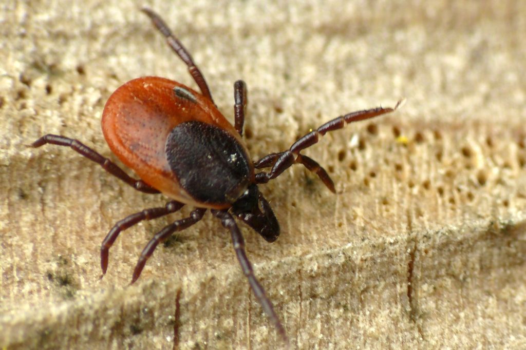
Ixodes ricinus (garrapata común)
Ixodes ricinus (castor bean tick)

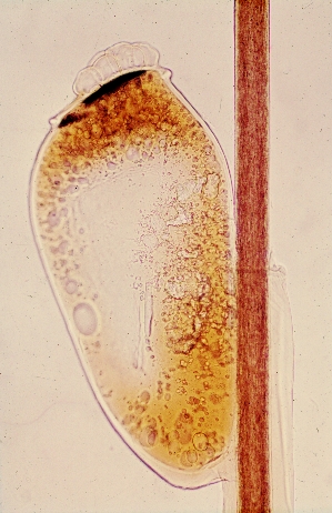
Liendre de piojo en un cabello
Louse egg in head hair
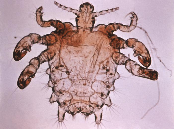
Pediculus humanus
Pediculus humanus
Pthirus pubis
Pthirus pubis
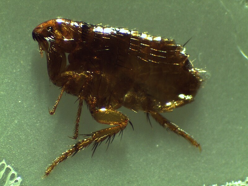
Pulga del gato (Ctenocephalides felis)
Cat flea (Ctenocephalides felis)
Cimex lectularius
Cimex lectularius

Videos


Crab louse (Pthirus pubis)


Human head louse (Pediculus humanus)


Human flea (Pulex irritans)

Questions and activities

1.- Draw the observed arthropods.

2.- Make a table or diagram of identification of the observed arthropods.

3.- Indicate the identifying characters of each arthropod observed, a small description and their parasitological importance.

4.- Search the Internet for images of arthropod parasites.

5.- Enter into the website Texas Agricultura & Life Sciences Agrilife Extension and search arthropod parasites.


Spanish version of this page


Parasitology
Leer más

Parasitic Helminthology


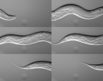
Nematod Caenorhabditis elegans in movement

The word helminth, worm, is commonly used in Parasitology. Two phylae of helminths are important in Parasitology: flatworms, and nematodes, roundworms (in old and alternative classifications appear in the Aschelminth phylum).

Platyhelminthes are flatworms, most are hermaphrodites and many are parasitic. Clonorchis sinensis affects more than 30 million people worldwide, especially in Asia. It is caused by consuming fresh or semi-processed fish or shellfish. The genus Opistorchis has a similar behavior, affecting, in addition to the human species, other mammals and fish.

Fasciola hepatica is a parasitic worm of the bile ducts and gallbladder of numerous species, including human. In livestock, it is one of the most widespread parasites in ruminants, especially cattle, goats and sheep, although it can also affect horses and lagomorphs. Its intermediate hosts are aquatic snails.

Schistosomiasis is produced by trematodes of the genus Schistosoma. It is not usually a serious illness, although it is incapacitating, mainly by the fever. Its biological cycle is typical of the trematodes, alternating in the same species of vertebrates and invertebrates.

Life cycle of Schistosoma
Taenia solium is transmitted to humans through uncooked or undercooked pork enters the human species generally through pork, while Taenia saginata does it for beef. Humans are generally infected for eating raw or undercooked beef which contains the infective larvae, called cysticerci.

The hydatid cyst is produced by Echinococcus granulosus. It is hydatidosis, which occurs in human species and certain animals, such as the dog and the sheep. It is produced by ingesting food contaminated by feces that contain the parasite. The disease can become deadly, as cysts can affect the liver, lungs, bones, kidneys or brain.

Hydatid cyst in lamb's liver
Nematodes form a large part of the planet's biomass and are critical to maintaining ecosystems. Parasite nematodes are a minority group of all nematodes.

Trichuriasis, also known as whipworm infection, is caused by the nematode Trichuris trichiura, which is characterized by gastrointestinal problems and diarrhea.

Trichinosis is produced by nematodes of the genus Trichinella through eating undercooked meat containing Trichinella cysts, most often this is pork but can also occur from bear and dog meat. This causes muscle pain, fever and gastrointestinal disorders. When the parasite reaches the heart, the consequences can be lethal or cause heart problems. If the infestation is massive it can cause death by the traumatic effect of the invasion of the tissues. For this reason, pork should not be consumed, if not analyzed with the trichinoscope, a microscope adapted for this purpose.

Trichinella spiralis, enrrollada en forma de espiral, de donde le viene el nombre
Trichinella spiralis, in muscle tissue
Strongyloides stercoralis produces strongyloidiasis. It is a common nematode in warm and humid areas. Strongyloidiasis is an important disease in immunocompromised persons and can cause a hyperinfection syndrome that can lead to death if untreated. It's produced by the contact of soils contaminated with feces.

Ancylostoma duodenale is a nematode that causes the hookworm infection, together with Necator americanus. It's one of the most frequent parasitosis in the world, which is contracted by the skin or orally in places of poor hygiene.

Ascaris lumbricoides causes ascariasis, in which the multiplication of helminths in the intestine causes health problems, such as diarrhea, pallor, malnutrition, etc. They may cause visceral damage, peritonitis, inflammation, appendicitis, intestinal obstruction, or migrate to other organs. The genus Toxocara affects several species, in addition to the human, such as canids and felids; In animals larvae migrate throughout the body and only affect the human species if the number of larvae is very high and the individual is immunosuppressed. Anisakis are parasites of fish that can affect the human species; Have proliferated in recent times, perhaps because of bad fishing practices (such as throwing guts into the sea) or changes in weather conditions.

The pinworm, also known as threadworm in the United Kingdom and Australasia or seatworm, is the specie Enterobius vermicularis, that causes pinworm infection, also known as enterobiasis, a disease spread all over the world, mainly affecting children under 12 years old, who spread the disease by scratching the anus by itching, extending the eggs of the parasite.

Filarias are parasitic nematodes that cause terror. Wuchereria bancrofti and Brugia malayi are transmitted through the mosquito bite, living in the lymphatic system and causing an enormous dilation of the tissues. Loa loa is transmitted through the sting of horseflies, living in the subcutaneous tissue. Onchocerca volvulus causes onchocerciasis, which is transmitted through the black fly, microfilariae move through the body and if they reach the eyes can cause blindness. Dranunculus medinensis (in the photo below) affects the subcutaneous tissues and causes dracunculiasis, is transmitted by ingesting contaminated water.
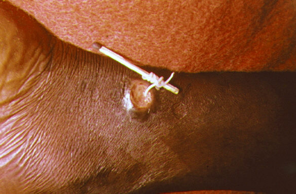
Dranunculus medinensis extraído a través de la piel
Dranunculus medinensis extracted through the skin

Acanthocephala is a phylum of parasitic worms known as acanthocephalans, thorny-headed worms, or spiny-headed worms, that possess a reversive proboscis with spines by which they adhere to the mucosa of the host. Macracanthorhynchus hirudinaceus have veterinary importance as it affects the pig.

The leeches are segmented worms that belong to the phylum Annelida and comprise the subclass Hirudinea. They live in the fresh water. Hirudo medicinalis is the common leech, which feeds on blood, distributed throughout much of Europe in fresh water. However, pollution, habitat destruction and hunting have almost completely eliminated their presence in the wild landscape.

Pentastomida are a group of parasitic crustaceans commonly known as tongue worms. All are obligate parasites with correspondingly degenerate anatomy. Linguatula serrata is a cosmopolitan zoonotic parasite, live in the nasopharyngeal region of mammals. Cats, dogs, foxes, and other carnivores are hosts of the parasite.


1. Platyhelminthes. General Characteristics and Classification

- Reading: Wikipedia. Flatworm
- Video: MicroStudent916. Helminths
- Video: Hark 07. Phylum Platyhelminthes


2. Clonorchis, Opisthorchis and Paragonimus

- Reading: Wikipedia. Clonorchis sinensis
- Reading: Wikipedia. Opistorchis viverrini
- Reading: Wikipedia. Opistorchis felineus
- Reading: Wikipedia. Paragonimus
- Video: TheLancetTV. Clonorchiasis
- Video: Miguel Vicente. Paragonimus westermani


3. Fasciola. Other trematoda

- Reading: Wikipedia. Fasciola hepática
- Reading: Wikipedia. Trematoda
- Video: MrChrisLeavitt. Sheep Liver Fluke Fasciola hepatica


4. Schistosoma

- Reading: Wikipedia. Schistosoma
- Reading: Wikipedia. Esquistosomiasis
- Video: Xistose.com. Schistosoma mansoni casal (Schistosoma mansoni couple)

5. Cestoda. General Characteristics and Classification

- Reading: Wikipedia. Cestoda
- Video: History Of Mankind Documentary. Worms - Documentaries - History Documentary


6. Pseudophyllidea: Diphyllobothrium latum

- Reading: Wikipedia. Diphyllobothrium
- Video: NEJMvideo. Live Diphyllobothrium Latum during Colonoscopy


7. Ciclophyllidea: Taenia

- Reading: Wikipedia. Taenia (genus)
- Reading: Wikipedia. Taenia solium
- Reading: Wikipedia. Taenia saginata
- Video: African Veterinary Information. Taenia solium cysticercosis: Life cycle, epidemiology and diagnosis


8. Echinococcus. Other cyclophyllidea

- Reading: Wikipedia. Echinococcus
- Reading: Wikipedia. Echinococcus granulosus
- Video: Neglected Tropical Diseases. Echinococcosis Documentry


9. Nematodes: General Characteristics and Classification

- Reading: Wikipedia. Nematoda
- Video: 7activestudio. Nematoda General Characters


10. Trichuris trichiura. Other trichuridae

- Reading: Wikipedia. Trichuris trichiura
- Reading: Wikipedia. Trichuridae
- Video: DrMurraSaca. Parasite in the Colon whipworm Trichuris trichiura


11. Trichinella

- Reading: Wikipedia. Trichinella
- Reading: Wikipedia. Trichinosis
- Video: Everything For You. Trichinell Spiralis (Complete Overview )

12. Strongyloides stercoralis

- Reading: Wikipedia. Stronglyoides stercoralis
- Reading: Wikipedia. Strongyloidiasis
- Video: Strongyloides stercoralis Larva life cycle


13. Uncinarias: Ancylostoma duodenale and Necator americanus

- Lectura: Wikipedia. Ancylostoma duodenale
- Lectura: Wikipedia. Necator americanus
- Video: Top Amazing. Parasite : Amazing found Ankylostome worm / Ancylostoma duodenal
- Video: BirdWhisperer46. Nematode - Necator americanus - Hook Worm


14. Ascaris lumbricoides. Toxocara, Anisakis. Other ascarids

- Reading: Wikipedia. Ascaris lumbricoides
- Reading: Wikipedia. Toxocariasis
- Reading: Wikipedia. Anisakis
- Reading: Wikipedia. Ascaris summ
- Reading: Wikipedia. Toxocara canis
- Video: A/G Geeks. Ascaris The Human Parasite
- Video: Sally Cantle. Toxocara Canis - The Dog Roundworm
- Video: Cristina Martínez. Anisakis Life Cycle (PARASITE project)


15. Enterobius vermicularis

- Reading: Wikipedia. Pinworm_(parasite)
- Video: Dr. Anisa Pal. ENTEROBIUS VERMICULARIS (gravid female) laying eggs-live video


16. Filarias: Wuchereria bancrofti, Brugia and Loa

- Reading: Wikipedia. Wuchereria bancrofti
- Reading: Wikipedia. Brugia malayi
- Reading: Wikipedia. Loa loa
- Video: Flexiguru. Elephantiasis/Filariasis


17. Onchocerca volvulus. Dracunculus medinensis. Other filarioidea

- Reading: Wikipedia. Onchocerca volvulus
- Reading: Wikipedia. Dracunculus medinensis
- Reading: Wikipedia. Filariasis
- Video: PublicResourceOrg. Filariasis


18. Acantocephala. Macracanthorhynchus hirudinaceus

- Reading: Wikipedia. Acantocephala
- Reading Wikipedia. Macracanthorhynchus hirudinaceus


19. Annelids leeches. Hirudo medicinalis

- Reading: Wikipedia. Hirudinea
- Reading: Wikipedia. Hirudo medicinalis
- Video: Dzikieswinie. Leech with cocoon - Hirudinea - Hirudo


20. Pentastómida. Linguatula serrata

- Reading: Wikipedia. Pentastomida
- Reading: Wikipedia. Linguatula serrata
- Video: Tamás Csordás. Linguatula serrata


Identification of helminth parasites. Virtual Lab


Resources for further learning

- Video: Boppolyscience. Platyhelminthes


Spanish version of this page


Parasitology
Leer más

Identification of helminth parasites. Virtual Lab

Scolex (head) of Taenia solium
The helminths or parasitic worms correspond to three types: Trematoda, or staves, are a class of phyllo Platyhelminthes (flatworms); Cestoda, are another class of the same phyla, and Nematode, another phyllo, tubular worms. Usually the helminths have a macroscopic size, measuring from 1 mm to 1 m (0,03937 in to 3,28 ft) , and even more.

The identification of the parasite is fundamental for the correct diagnosis of the parasitosis. This requires training in microscopic techniques staining and identification.


Background

- Reading: Para-Site. Helminth Parasites


Procedure

The microscopic identification techniques of parasitic helminths are complex and require specialized training.

This experiment focuses on the identification of parasitic helminths by microscopic and macroscopic observation, not in the manipulation of samples, nor in the microscopic techniques of preparation and staining. For this, a series of parasitic helminths will be observed.

For the identification of cestodes:

- Reading: M. Arcari; A. Baxendine; C. E. Bennett. Guide to Parasitology. The Cestodes

For the identification of nematodes:

- Reading: M. Arcari; A. Baxendine; C. E. Bennett. Guide to Parasitology. The Nematodes

For the identification of trematodes:

- Reading: M. Arcari; A. Baxendine; C. E. Bennett. Guide to Parasitology. The Trematodes


Ways to perform the experiment

1. In Laboratory

The laboratory that performs parasitic helminthology experiments and tests, must have a supply of samples, adequate instruments and staff training. If this is not possible, the microscopic preparations can be purchased. The microscopic preparations of helminth parasites usually cost about 8-10 dollars each. If macroscopic samples of helminths are not available, a good and alternative option is to make visits to museums of Natural History.


2. In Home Lab

In principle, the experiment is not dangerous if carried out with commercial microscopic preparations. For the experiment it is necessary to have a microscope something better than a toy. There are possibilities for setting up a low cost home microscopy lab (in spanish).

If macroscopic samples of helminths are not available, a good alternative option is to make visits to museums of Natural History.


3. Virtually

For this purpose, microscopic images of parasitic helminths can be made in the same post in the next section.


Images of helminth parasites

Click on the image to enlarge.

Fasciola hepatica
Fasciola hepatica

Schistosoma mansoni
Schistosoma mansoni

Proglótidos de Diphyllobothrium latum
Proglottids of Diphyllobothrium latum

Taenia saginata
Taenia saginata

Trichinella spiralis en tejido muscular
Trichinella spiralis in muscle tissue

Huevos de Ancylostoma duodenale
Eggs of Ancylostoma duodenale

Huevo de Ascaris lumbricoides
Egg of Ascaris lumbricoides

Ascaris lumbricoides

Toxocara canis
Toxocara canis

Anisakis en arenques
Anisakis in herring
Huevo de Enterobius vermicularis
Egg of Enterobius vermicularis
Videos:


Removing by surgery of Ascaris lumbricoides


Removing a filarial worm in a eye

Questions and activities


1.- Draw the observed helminths.

2.- Make a table or diagram of identification of the observed helminths.

3.- Indicate the identifying characters of each helminth observed, a small description and their parasitological importance.

4.- Search the Internet for images of helminth parasites.

5.- Enter into the website Global Atlas of Helminth Infections, to Maps and observe in the maps the distribution of the parasitic diseases for helminths, for country or for type of parasite.


Resources for further learning
Leer más

Parasitic Protozoology

Quiste de Entamoeba histolytica
Entamoeba histolytica cyst
Protozoa are microscopic eukaryotic organisms. Formerly they were classified as animals (Animalia kingdom) and today it is known that they have some characteristics that they share with the vegetables, being a very diverse group, with relation of remote kinship. There are classifications that encompass them in Protist, with different phyla, being a paraphyletic group. Others create for them a eukaryotic kingdom Protozoa. Parasitic protozoa are a minority and need a host. Sometimes to overcome disfavorable conditions become cyst.

Amoebas are protozoans that move by movement accomplished by protrusion of cytoplasm involving the formation of pseudopodia (amoeboid movement). The best known parasite is Entamoeba histolytica, which parasites dogs and people. It causes amoebic dysentery, feeding on the bolus of food and intestinal bacteria, even the intracellular fluids of the cells it destroys.

Intestinal ulcer caused by Entamoeba histolytica
Giardia lamblia, intestinalis or duodenalis is a flagellate protozoan that lacks certain organelles such as mitochondria and the Golgi apparatus and produces intestinal conditions that can become serious. It was the first microscopic parasite found in the human species by Anton van Leeuwenhoek when he observed under the microscope fecal matter, but at that moment nothing was known about protozoa.

The protozoan Trichomonas vaginalis causes trichomoniasis, a sexually transmitted disease that in some populations of industrialized countries has an incidence of 70%. Most people infected do not have any symptoms. Symptoms experienced include pain, burning or itching in the penis, urethra, or vagina.

Leishmaniasis is a disease caused by the protozoan Leishmania, transmitted through mosquitoes. It is a disease that affects dogs and humans (zoonoses). Some species of Leishmania cause severe ulcers on the skin leaving permanent scars.

Trypanosoma causes african trypanosomiasis, also known as sleeping sickness. The trypanosomes are passed in the saliva of the tsetse fly. One to three weeks after the bite, in the first stage of the disease, there are headaches, fevers, and joint pains. Weeks to months later the second stage begins with confusion, poor coordination, and trouble sleeping. Without treatment it typically results in death.

Apicomplexa is a group of endoparasite protists that penetrate the inside of cells. Cryptosporidium causes a diarreic disease; Toxoplasma the disease toxoplasmosis; Plasmodium malaria; and Babesia infect the blood and cause a hemolytic disease known as babesiosis.

Toxoplasmosis is an infectious disease caused by the protozoan Toxoplasma gondii, an obligate intracellular parasite. It is a zoonosis that is transmitted from animals to humans through different routes of infection, being definitive hosts the cat and six other species of felines, but the most important route of infection are contaminated food. It has a high incidence in Africa and America, with 80% being asymptomatic infestations, and in other cases chronic.

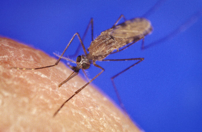
Malaria is a disease caused by the genus Plasmodium, through the mosquitoes of the genus Anopheles. It has a high incidence worldwide, causing two to three million deaths a year.

Anopheles gambiae, transmisor de la malaria
Anopheles gambiae, transmitter of malaria
Babesiosis is caused by the protozoan Babesia, which attacks the red blood cells and transmitted by ticks. In the cattle causes the denominated Texas cattle fever.


1. Protozoans. General characters and classification

- Reading: Wikipedia. Protozoa
- Video: Mr. Rawlings Biology. The World of the Protozoa


2.- Amoebiasis. Entamoeba histolytica. Free-living Amoebozoa infection

- Reading: Wikipedia. Amoebiasis
- Reading: Wikipedia. Entamoeba histolytica
- Reading: Wikipedia. Free-living Amoebozoa infection
- Video: DrER.tv. Amebiasis


3. Intestinal Flagellates. Giardia lamblia

- Reading: Wikipedia. Flagellate
- Reading: Wikipedia. Giardia lamblia
- Presentation: Biology Teaching Centre. Flagellates (pdf)


4. Trichomonanida. Trichomonas vaginalis

- Reading: Wikipedia. Trichomonanida
- Reading: Wikipedia. Trichomonas vaginalis


5. Leishmania

- Reading: Wikipedia. Leishmania
- Video: Let's Learn About Bugs. What is Leishmaniasis? An introduction and overview


6. Trypanosoma

- Reading: Wikipedia. Trypanosoma
- Video: videosINBEB. Animated life cycle of T. cruzi in the human host


7. Apicomplexa. General characters and classification

- Reading: Wikipedia. Apicomplexa
- Vídeo: iBiology. David Roos (U Penn) Part 1: Biology of Apicomplexan Parasites
- Vídeo: iBiology. David Roos (U Penn) Part 2: The apicomplexan plastid


8. Cryptosporidium

- Reading: Wikipedia. Cryptosporidium
- Video: Paul Cochrane. Cryptosporidium: What You Should Know


9. Cyclospora, Cystoisospora, Sarcocystis and Toxoplasma

- Reading: Wikipedia. Coccidia
- Reading: Wikipedia. Cyclospora cayetanensis
- Reading: Wikipedia. Cystoisospora belli
- Reading: Wikipedia. Sarcocystis
- Reading: Wikipedia. Toxoplasma gondii
- Video: Maddie's Fund Education. Coccidia in a Shelter Setting


10. Plasmodium and Babesia

- Reading: Wikipedia. Plasmodium
- Reading: Wikipedia. Babesia
- Video: Armando Hasudungan. Malaria - Plasmodium


Identification of protozoans parasites. Virtual Lab


Resources for further learning

- Lectura: Wikipedia. Acanthamoeba
- Lectura: Wikipedia. Balamuthia mandrillaris
- Lectura: Wikipedia. Naegleria


Spanish version of this page


Parasitology
Leer más

Identification of protozoan parasites. Virtual Lab

Plasmodium falciparum
Plasmodium falciparum, protozoan that causes malaria in humans
Some of the protozoa, these microscopic protists, who share characteristics of plants and animals and live in water, are a serious problem for human and animal health, since many of them are parasites and cause very serious diseases.

According to the World Health Organization (WHO), in the year 2015 there were about 214,000,000 cases of malaria, a disease caused by the protozoan Plasmodium, which killed 438,000 people. Leishmaniasis, caused by the protozoan Leishmania is a disease that affects dogs and humans, causes severe ulcers on the skin leaving scars for life. The trichomoniasis, a sexually transmitted disease, is caused by the protozoan Trichomonas vaginalis. Some sources estimate an incidence of 70% in populations of developed countries, with no symptoms or sporadic symptoms such as itching or pain in sexual intercourse. These are just some examples of the importance of parasites in human health. And this without mentioning the importance in animal health, both in the animals that surround us, and in the livestock economy.

The identification of the parasite is fundamental for the correct diagnosis of the parasitosis. This requires a training in microscopic staining and identification techniques, and in some cases additional tests, such as the ELISA test.


Background

- Reading: Austin Community College. Medical Parasitology. Lab Manual ( LABORATORY EXERCISE 3:  Identification of Parasites)


Procedure

Microscopic identification techniques for protozoan parasites are complex and require specialized training.

This experiment will focus, not on the manipulation of the samples, nor on the microscopic techniques of preparation and staining, but on the identification of protozoan parasites through microscopic observation. For this, a series of intestinal protozoan parasites will be observed under the microscope.

- Reading: M. Arcari, A. Baxendine and C. E. Bennett. Diagnosing Medical Parasites Through Coprological Techniques: Volume 1 The Amoebae  See BelowVolume 2. The Ciliates, Coccidia and MicrosporidiaVolume 3. The Flagellates


Ways to perform the experiment

1. In Laboratory

The laboratory that performs experiments and tests of parasitic protozoology, must have a supply of samples, adequate instruments and staff training. If this is not possible, microscopy preparations can be acquired. One microscopy preparation of parasitic protozoa usually cost about 8-10 $.

2. In Home Lab

In principle, the experiment is not dangerous if carried out with commercial microscopic preparations. For the experiment it is necessary to have a microscope something better than a toy. There are possibilities for setting up a low cost home microscopy lab (in spanish).

3. Virtually

For this purpose, microscopic images of parasitic protozoa can be made in the same post in the next section.


Microscopic images of protozoan parasites

Click on the image to enlarge.

Entamoeba histolytica
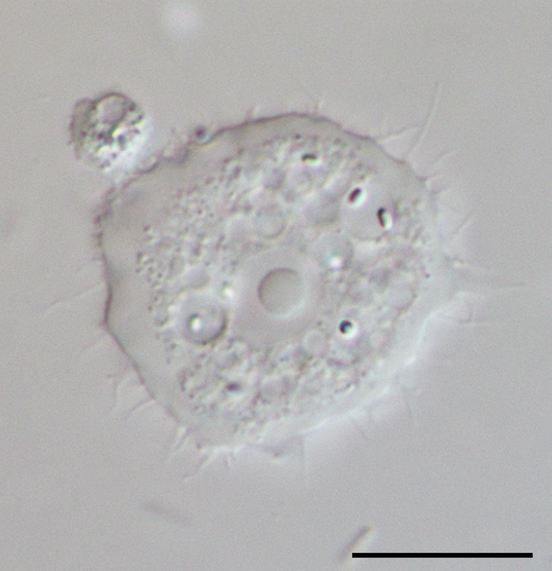
Acanthamoeba
Acanthamoeba

Trichomonas vaginales (tinción Giemsa)
Trichomonas vaginalis

Tripanosoma cruzi
Tripanosoma cruzi

Plasmodium falciparum

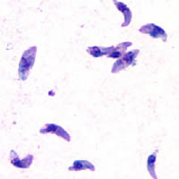
Toxoplasma gondii
Toxoplasma gondii

Leishmania donovani
Leishmania donovani in bone marrow cells (in the center)

Babesia
Babesia


Giardia lamblia


Pentatrichomonas hominis, fresh analysis of human feces


Trichomonas vaginalis, thawed after freezing


Questions and activities

1.- Draw the observed protozoa.

2.- Make a table or diagram of identification of the observed protozoa.

3.- Indicate the identifying characters of each protozoan observed, a small description and their parasitological importance.

4.- Search the Internet for images of protozoan parasites.

5.- Enter into the website Protist Image Data, access the database of protists, identify protozoan parasites and access their images and data (the website has some years and there are several broken links).


Resources for further learning

- Reading: World Health Organization. Basic laboratory methods in medical parasitology (pdf)


Spanish version of this page


Parasitology
Leer más

The Science of Parasitology

Black fly (Simulium yahense)

Parasitology is the part of biology that studies parasitism, and their field of study and research are parasitic organisms and their relationship with their hosts and the environment. This science is related intimately with Zoology, Ecology and Microbiology. Parasitology has important implications in medicine, pharmacy and veterinary, constituting the medical parasitology and veterinary parasitology, applied sciences to health, that besides studying parasitism, too investigates the solutions.

Parasitism is a form of symbiosis or common life of living beings, is its dark side, the opposite of mutualism, in which both species are favored, because parasitism involves exploitation of species. Its a part of the evolution, since both species evolve over time. There are degrees, from facultative parasites to obligate parasites, which can not survive by themselves.

The parasite needs a host and overcome what can separate it. Ectoparasites or external parasites, animals and plants, have it easier than endoparasites or internal parasites, because the most common route of entry is the digestive, as they have to overcome barriers of the immune system, temperature, osmotic pressure, etc. This whole process of adaptation arise in invertebrates living in hostile systems.

Although the parasite is harmful, isn't interested in being lethal to the host, as their way of life and livelihood would end, must be in balance, which is reached by coevolution, the two species have evolved in an interaction. It is estimated that parasitism already exist in the Precambrian, some 1,100 million years ago.

The parasite must have certain characteristics that facilitate their way of life: ease of reproduction, organs for fixation, loss of sensory organs (not to waste energy), large egg production, lack of pigments, adapting their cycles to the host, etc. Moreover, the host may be final or intermediary. The parasite can have a life cycle, having an internal or external phase, or only one; or have the cycle is directly or indirectly through one or more intermediate hosts.

Among the major groups of parasites are protozoa; helminth, worms, flatworms (flattened flukes and tapeworms) and aschelminthes (cylindrical, the most important nematodes); and arthropods. But there are also other parasites groups such as fungi (Cordyceps, parasite of insects), viruses, or vertebrates, like the cuckoo, a bird.

The identification techniques of endoparasites may be direct (liquid stool samples) or indirect (ELISA test). The identification of hemoparasites and intercellular parasites, found in the blood or tissues may be direct (such as blood smear or sample analysis) or indirect (such as serum antibodies). The identification of arthropods are usually direct, by observation with a magnifying glass or microscope.


1. Parasitology

- Reading: Wikipedia. Parasitology


2. Interspecific interactions and parasitism. Origin of parasitism

- Reading: Wikipedia. Parasitism


3. Main classes of parasites

- Reading: CDC. About Parasites


4. The parasitology lab: diagnosis approaches and methods

- Reading: Hindawi. Diagnosis of Parasitic Diseases: Old and New Approaches


Resources for further learning

Leer más